Document Type : Original Research Article
Authors
Al-Farahidi University, Pharmacy College, Department of Pharmaceutics, Baghdad, Iraq
Abstract
Electrospinning is used to prepare fine fibers mats from polyvinyl alcohol (PVA) solution that contains diclofenac potassium (DP) which is used in the treatment of local pain on the affected area to reduce the inflammation. The solution and process parameters (i.e. solution concentration, applied voltage current, flow rate, and needle-collector distance) affect the morphology of the fibers; which was studied using SEM to obtain fibers with diameter within the nano-size range. Generally, the average nanofibers diameter that obtained from SEM images are 1115 nm and 1864 nm for 10% PVA/DP and 15% PVA/DP, respectively, which indicate that, the diameter of nanofiber diameter increases with increasing the concentration of PVA at constant applied voltage current 30 Kv, flow rate of 0.5 mL/hour, and needle-collector distance 8 cm.
Graphical Abstract
Keywords
Main Subjects
Introduction
Electrospinning is a technique used to produce nanofibers from a polymer solution. The process involves applying a high voltage current to a polymer solution, which creates an electrical charged jet of fluid that is stretched and elongated by the electric field, forming very fine fibers. Then the fibers collected on a grounded collector, resulting in a non-woven mat or membrane of nanofibers [1].
Electrospinning is a versatile technique that can be used to produce fibers from a wide range of materials, including synthetic and natural polymers, and has numerous applications in the medical fields such as biomedical engineering, tissue engineering, drug delivery, filtration, energy storage, and environmental remediation [2].
One of the major advantages of electrospinning is that it can produce fibers with diameters in the nanometer range, which makes them useful in a wide range of applications where a high surface area to volume ratio is desirable [3]. However, the technique does have some challenges, such as controlling fiber morphology and uniformity; thus researchers are working to address through the development of new equipment and methods [4].
The fibers exhibit several interesting characteristics, e.g., high surface area to volume ratio, surface flexibility, and high porosity. These unique features render electrospun fiber as good candidates for use in various pharmaceutical and biomedical applications such as tissue engineering scaffolds and drug delivery systems [5]. The process of preparing electrospun fibers involves using an electrospinning unit, where a high voltage current is applied to a polymer solution dispensed from a syringe needle. This process creates ultrafine fibers by applying a high electrical potential from an emitting electrode of a high-voltage power supply to the polymer liquid. The distance between the conductive nozzle and the grounded collector is crucial in facilitating the fabrication of these fibers through electrospinning [6]. The repulsion force between charges of the same polarity generated in the polymer liquid destabilizes the partially-spherical droplet of the polymer liquid located at the tip of the nozzle to finally form a droplet of a conical shape (i.e. the charged jet).
The main components of an electrospinning device include
(1) High-voltage power supply: This component provides the high voltage necessary to create an electric field between the spinneret and the collector.
(2) Spinneret: This is a small, needle-like device that is used to deliver the polymer solution to the electric field.
(3) Polymer solution delivery system: The polymer solution is delivered to the spinneret through a syringe pump or similar system.
(4) Collector: This is a grounded plate or drum that collects the nanofibers as they are electrospun.
(5) Electrode: This is a conductive material, usually a metal wire or plate that is used to create the electric field between the spinneret and the collector.
(6) Enclosure: An enclosed chamber is used to control the environment during the electrospinning process, such as temperature and humidity.
(7) Syringe: This is used to hold the polymer solution before it is delivered to the spinneret [7].
Overall, an electrospinning device can vary in complexity and design depending on the specific application and requirements of the process. The electrospinning device is displayed in Figure 1.

Figure 1. The Components of electrospinning device [7]

Figure 2. The chemical structure of PVA
The electrospun fibers are produced by use natural and synthetic which developed as the matrix materials for delivery of drugs; synthetic polymers are used as Drug Delivery Systems (DDS) such as PVA. The structure of PVA is depicted in Figure 2.
Polyvinyl alcohol (PVA) is a water-soluble synthetic polymer that is commonly used in electrospinning to produce nanofibers. PVA Structure or polyvinyl alcohol structure vinyl acetate formula is H3C-CO2-CH=CH2. PVA is not prepared by direct polymerization of vinyl acetate [8]. Instead, it is prepared by hydrolysis of polyvinyl acetate (PVAc) through an alcohol (generally methanol) in presence of an alkaline catalyst. PVA have several properties that make it a good candidate for electrospinning applications, including:
Water solubility
PVA is highly soluble in water, which makes it easy to prepare a polymer solution for electrospinning. Biocompatibility of PVA is non-toxic and biocompatible, which makes it suitable for a variety of biomedical applications [9]. A mechanical property has good tensile strength and flexibility, which makes it ideal for producing strong and durable nanofibers. Chemical stability is resistant to chemical and biological degradation, which makes it suitable for applications where long-term stability is important. Electrospinnability can be electrospun into nanofibers with a range of diameters, making it useful for a variety of applications. PVA-based electrospun nanofibers have been used in various applications, including drug delivery, tissue engineering, and filtration. PVA is also often used as a component in blend electrospinning with other polymers to improve the mechanical properties of the resulting nanofibers [10].
Diclofenac potassium is a nonsteroidal anti-inflammatory drug (NSAID) that is commonly used to treat pain, inflammation, and fever. It is often used to relieve symptoms associated with conditions such as osteoarthritis, rheumatoid arthritis, and ankylosing spondylitis [11].
In recent years, there has been growing interest in using diclofenac potassium in electrospun nanofibers for external use, as this method can enhance the drug's effectiveness and provide targeted delivery to affected areas. Electrospinning is a process that involves the application of an electrical field to a polymer solution, resulting in the formation of nanofibers that can be used to encapsulate drugs. When diclofenac potassium is incorporated into electrospun nanofibers, it can be applied topically to provide localized pain relief and anti-inflammatory effects. The nanofibers can also improve the drug's bioavailability, as they provide a larger surface area for absorption and can release the drug over an extended period [12].
Overall, the use of diclofenac potassium in electrospun nanofibers represents an exciting development in drug delivery technology, offering a potentially effective and targeted approach to managing pain and inflammation. The chemical structure of diclofenac potassium is demonstrated in Figure 3.

Figure 3. The chemical structure of diclofenac potassium
Experimental
Materials and Methods
The materials used in this study included Polyvinyl alcohol (Hainan Huarong Chemical Co. Ltd.; India, Mw 120.000 KD), diclofenac potassium (Xa-Bc-Biotech Co.,Ltd; China); Glutaraldehyde (GA) 25% (Acuro Organics Limited; India); Phosphate Buffer Solution 7 (CPAChem; Bulgaria), Disodium Hydrogen Sulphate and Potassium Dihydrogen Phosphate (Himedia laboratories; India); and Deionized water (Beea Alrafidain; Iraq); all chemicals were purchased from Al-Noor medical store for chemicals. The remaining materials used are available as laboratory grade.
Preparation phosphate buffer solution pH 5.5
Solution I:
13.61 g of potassium dihydrogen phosphate was dissolved in water and diluted to 1000mL with the same solvent.
Solution II:
35.81 g of disodium hydrogen phosphate was dissolved in water and diluted to 1000mL with the same solvent. 96.4 mL of solution I and 3.6 ml of solution II were mixed [13].
Preparation of formulas
There are the formulas to prepare the electrospun solutions of diclofenac potassium for each PVA concentration:
For PVA 10%:
Diclofenac potassium: 100 mg/mL (to be dissolved in PBS pH 5.5 (10%) w/v)
PVA (10% w/v): 1 g/100 mL solvent mixture
Solvent: 9 mL of a 50:50 mixture of water and ethanol
For PVA 15%:
Diclofenac potassium: 100 mg/mL (to be dissolved in PBS pH 5.5 (10%) w/v)
PVA (15% w/v): 1.5 g/100mL solvent mixture
Solvent: 8.5 mL of a 50:50 mixture of water and ethanol
To prepare the solutions, you can follow these steps:
Weigh out the required amount of diclofenac potassium and PVA separately.
Add the PVA to the solvent and stir until it dissolves completely. If the solution is too thick, you can heat it slightly to facilitate the dissolution process.
Add the diclofenac potassium to the PVA solution and stir until it dissolves completely.
The electrospun solution is now ready to use [14]; the formulas of electrospun solution is indicated in Table 1.
Table 1. Formulas of electrospun nanofibers solution
|
Formulas |
DP w/v |
PVA w/v |
|
F1 |
100 mg/mL |
10% |
|
F2 |
100 mg/mL |
15% |
The actual concentration of diclofenac potassium in the electrospun fibers will depend on various factors, such as the flow rate of the electrospinning solution and the parameters used during electrospinning.
Process of electrospinning nanofibers (DP/PVA)
The most common method of blending diclofenac potassium (DP) with polyvinyl alcohol (PVA) to prepare electrospun nanofiber solution is the solution blending method. This method involves dissolving both DP and PVA in a solvent mixture of water and ethanol. The DP and PVA solutions are then mixed together and stirred until they are homogenous.
After the DP and PVA solutions are homogenous, the resulting solution can be used for electrospinning to form nanofibers. During electrospinning, the solution is loaded into a syringe of (needle size of 23G) with keeping the needle-collector distance to about 8cm and then subjected to a high voltage of (30 Kv) [15]. The rate of flow is controlled using HPLC pump of (0.5 mL/hour); which creates a charged jet that is extruded through a small needle onto a collector cylinder. The charged jet undergoes a whipping motion that causes it to elongate and form nanofibers as it travels from the needle to the collector. The prepared nanofibers are treated with Glutaraldehyde (GA) 25% vapor to about 2-hour; left until dried at room temperature for 8-hour. Finally removing the prepared nanofibers from the collector with care to be ready for the next step which represented in the scanning the product with SEM to determine the fibers diameter for each formula [16].
It is important to note that the parameters used during the electrospinning process, such as voltage, flow rate, and distance between the needle and collector can affect the morphology, diameter, and alignment of the resulting nanofibers. Therefore, optimizing these parameters is crucial to obtain the desired properties of the nanofibers for a specific application [17].
Scanning electron microscopyTop of Form
The structural analysis of SEM analysis of diclofenac potassium/PVA formulations (using a Thermo-Fisher Scientific electron microscope, Japan) typically involves imaging the surface of the drug particles and the PVA matrix. This can provide information on the particle size and distribution, as well as the morphology of the particles and their interaction with the PVA matrix.
Overall, SEM can be a useful tool for investigating the microstructure and morphology of diclofenac potassium/PVA formulations and can provide valuable insights into their properties and behavior. However, the specific details of any study would depend on the research question and methodology used.
Results and Discussion
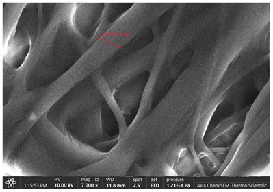
The scanning electron microscope (SEM) images of diclofenac potassium and PVA with different nanofiber diameters provide valuable information about the morphology and structure of the electrospun fibers. In particular, the comparison between 10% and 15% PVA solutions with diclofenac potassium is of interest. The SEM images show that the electrospun fibers have a uniform morphology and smooth surface with no visible defects. The average diameter of the nanofibers is about 1115 nm for the 10% PVA/DP solution and 1864 nm for the 15% PVA/DP solution, indicating that the fiber diameter increases with the increase in PVA concentration.
In the case of diclofenac potassium incorporated nanofibers, the SEM images show that the drug is uniformly dispersed within the fibers, without any agglomeration or clustering. This indicates that the drug is well incorporated within the polymer matrix, which is essential for controlled drug release applications.
Overall, the SEM results suggest that the electrospinning process is successful in producing uniform and smooth nanofibers with a diameter that can be controlled by adjusting the PVA concentration. The incorporation of diclofenac potassium within the fibers is also successful, without any significant changes in the fiber morphology or drug distribution. The resultant images of (SEM) are demonstrated in Figures 4 and 5, respectively.

Figure 4. Scanning electron microscope image of F1

Figure 5. Scanning electron microscope image of F2
Conclusion
The scanning electron microscope (SEM) images offer significant insights into the morphology and structure of electrospun nanofibers incorporating diclofenac potassium and PVA. Our findings indicate that as the PVA concentration increases, there is a corresponding increase in the average diameter of these fibers. This trend highlights the considerable potential of these nanofibers for drug delivery applications.
Disclosure Statement
No potential conflict of interest was reported by the authors.
Funding
This study was funded by Payame Noor University (PNU) Research Council.
Authors' Contributions
All authors contributed to data analysis, drafting, and revising of the article and agreed to be responsible for all the aspects of this work.
Orcid
Ahmad AB Yosef Kinani
https://orcid.org/0000-0001-7938-4294
Mohammed H. Al-Bayati
https://orcid.org/0009-0001-9122-6931
How to cite this manuscript: Ahmad AB Yosef Kinani, Mohammed H. Al-Bayati. Green Preparation of Diclofenac Potassium via Electrospun Nanofibers Based on Synthetic Polymer. Asian Journal of Green Chemistry, 8(4) 2024, 373-381. DOI: 10.48309/AJGC.2024.449737.1491


